 Add to Chrome
 Add to Firefox
 Add to Edge
Toxicity Assessment in Preclinical Histopathology via Class-Aware Mahalanobis Distance for Known and Novel Anomalies
Feb 02, 2026Drug-induced toxicity remains a leading cause of failure in preclinical development and early clinical trials. Detecting adverse effects at an early stage is critical to reduce attrition and accelerate the development of safe medicines. Histopathological evaluation remains the gold standard for toxicity assessment, but it relies heavily on expert pathologists, creating a bottleneck for large-scale screening. To address this challenge, we introduce an AI-based anomaly detection framework for histopathological whole-slide images (WSIs) in rodent livers from toxicology studies. The system identifies healthy tissue and known pathologies (anomalies) for which training data is available. In addition, it can detect rare pathologies without training data as out-of-distribution (OOD) findings. We generate a novel dataset of pixelwise annotations of healthy tissue and known pathologies and use this data to fine-tune a pre-trained Vision Transformer (DINOv2) via Low-Rank Adaptation (LoRA) in order to do tissue segmentation. Finally, we extract features for OOD detection using the Mahalanobis distance. To better account for class-dependent variability in histological data, we propose the use of class-specific thresholds. We optimize the thresholds using the mean of the false negative and false positive rates, resulting in only 0.16\% of pathological tissue classified as healthy and 0.35\% of healthy tissue classified as pathological. Applied to mouse liver WSIs with known toxicological findings, the framework accurately detects anomalies, including rare OOD morphologies. This work demonstrates the potential of AI-driven histopathology to support preclinical workflows, reduce late-stage failures, and improve efficiency in drug development.
Learning image representations for anomaly detection: application to discovery of histological alterations in drug development
Oct 14, 2022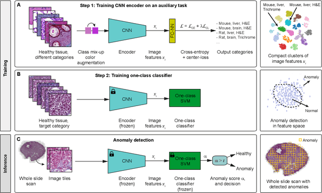
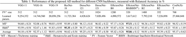
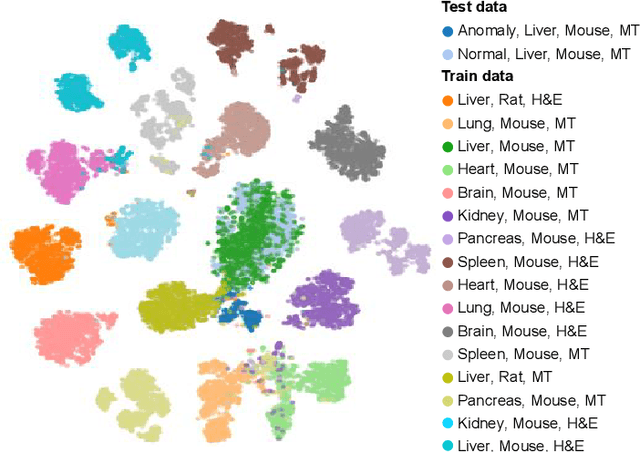
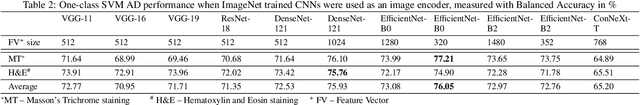
We present a system for anomaly detection in histopathological images. In histology, normal samples are usually abundant, whereas anomalous (pathological) cases are scarce or not available. Under such settings, one-class classifiers trained on healthy data can detect out-of-distribution anomalous samples. Such approaches combined with pre-trained Convolutional Neural Network (CNN) representations of images were previously employed for anomaly detection (AD). However, pre-trained off-the-shelf CNN representations may not be sensitive to abnormal conditions in tissues, while natural variations of healthy tissue may result in distant representations. To adapt representations to relevant details in healthy tissue we propose training a CNN on an auxiliary task that discriminates healthy tissue of different species, organs, and staining reagents. Almost no additional labeling workload is required, since healthy samples come automatically with aforementioned labels. During training we enforce compact image representations with a center-loss term, which further improves representations for AD. The proposed system outperforms established AD methods on a published dataset of liver anomalies. Moreover, it provided comparable results to conventional methods specifically tailored for quantification of liver anomalies. We show that our approach can be used for toxicity assessment of candidate drugs at early development stages and thereby may reduce expensive late-stage drug attrition.